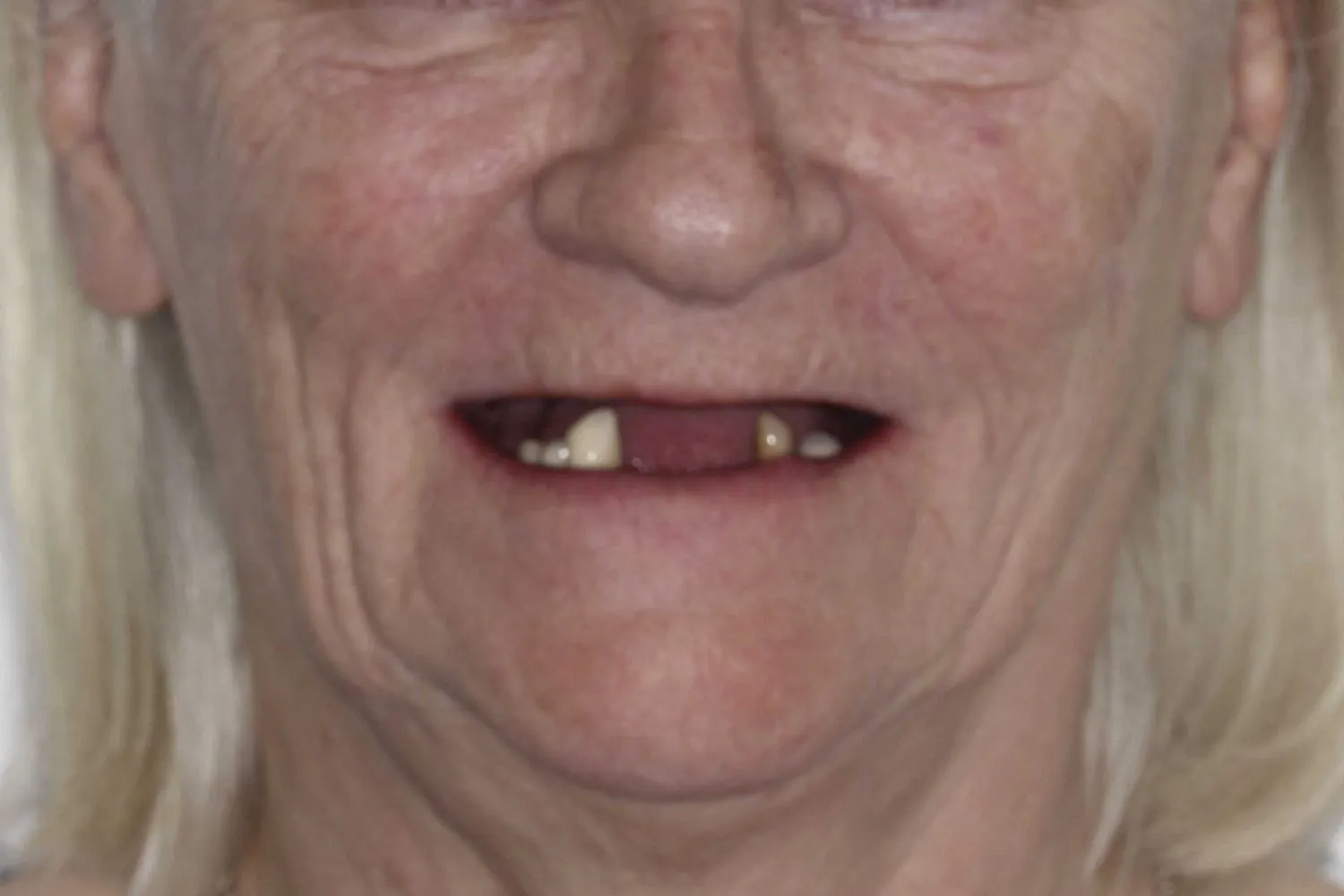
Mrs. G.H. attended our practice with aesthetic concerns. She had done composite fillings on a central incisor and a ceramic crown on the lateral. The lateral was an implant placed as a teenager and the anterior work replicated her teenage dentition. This was causing anxiety issues when interacting socially.
After an initial aesthetic, clinical and radiological evaluation, the patient was informed regarding the performance of the existing fillings and expected outcome.
The patient was not happy with the size and shape of the existing central and lateral, so a decision was made to replace the existing restorations of the anterior upper right central incisor and lateral using ceramic indirect restorations.
The planning stage included study models, photographs and shade records were taken. A difference in available space was detected. The lateral to be restored had to be bigger than the contralateral. A decision was made to create and illusion of symmetry by carefully placing the transition lines1.
Impressions were made and a digital wax-up of the lateral and central was presented to the patient. The patient approved the shape and size2.
Removal of the existing central incisor composite restoration was performed and removal of 0,5mm to 1mm of tooth structure for a veneer was done. The tooth surface was polished using discs and the margins perfected with chisels. Silicon index guides were used to check if even space was available for the ceramic restorations3.
The stump shade was recorded using eLAb protocol and impressions were made after retraction cord was placed on the sulcus.
To optimise aesthetics and adhesion to dentin and enamel, an adhesive cementation technique using Lithium disilicate veneer was chosen for the central. A gold Atlantis abutment with a zirconia crown was chosen for the lateral.
A temporary veneer was made using self-curing acrylic.
Once the lab-made restorations were ready, thickness was measured on several locations and photographic records taken to check margins and aesthetics.